
Clinical Significance
Despite their success and popularity, prosthetic AVGs are fraught with multiple modes of failure.
Modes of AVG graft failure include: thrombosis, infection, weeping of serous fluid, aneurysm formation, and traumatic degradation of the graft material. Many of these complications result from improper needle cannulation and graft handling, but can also be attributed to inferior material and construction of current AVGs. Graft degradation can be significantly accelerated by inadvertent punctures to the posterior or sidewall of the graft. These unintentional punctures can result in a peri-graft hematoma or pseudoaneurysm formation, which can lead to graft bleeding, thrombosis, infection, and failure.
Multiple modes of failure from AVGs
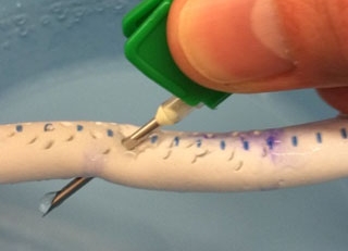
Accidental needle penetration.
Standard 16 ga dialysis needle easily penetrates the back wall of a standard dialysis graft.

Hematoma following needle injury.
Extremely large (about the size of a mini football) hematoma (collection of blood) beneath the skin of patient who suffered a needle injury during an attempted dialysis treatment.

Infection following sheath injury.
Infected pseudoaneurysm (outpouching from graft with live blood flow) following a sheath injury during a common dialysis graft angiogram and thrombectomy (declot procedure).
Graft degradation from repetitive needle puncture
Overly aggressive graft compression, to provide hemostasis following needle withdrawal, can result in graft thrombosis and failure. Repetitive cannulation and coring of the AVG material results in graft degradation, pseudoaneurysm formation, and graft rupture.
Over time, pseudoaneurysms develop mural thrombi, which may cause either thrombosis or infection of the graft. Continued growth of pseudoaneurysms may result in skin excoriation, infection, rupture, hemorrhage, exsanguination, and death.
These complications result in increased pain, disability, surgical intervention, and cost associated with the care of patients with end stage renal disease.
Example of dialysis graft degradation from repetitive needle puncture, and corresponding angiogram (below).

